


_GFAP(594)_40X-2%20merge.jpg)
_4Rtau(594)_40X-4%20merge.jpg)

Comprehensive Red Fluorescence Microscopy Image Gallery
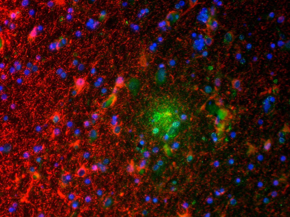
This gallery presents a diverse collection of fluorescence microscopy images, captured using various techniques and settings. The images were taken with a Red Fluorescence Microscope and stained with a range of fluorophores, including DAPI, GFP, Texas Red, Cy3, and Cy5, to visualize specific cellular components. A variety of objectives were used to capture these images at different magnifications, including 4X, 10X, 20X, and 40X, allowing for both a broad view and detailed examination of the samples. The sample thickness also varies from 5µm to 30µm, providing insights into different tissue and cell structures. This collection highlights the versatility of fluorescence imaging in revealing cellular and tissue morphology across multiple scales.


-20X%204000%203000.jpg)
.jpg)
LE.AM Solution
Tel. 82-31-434-2710
Hwasung-Si, Gyeong-gi Do,
South Korea.